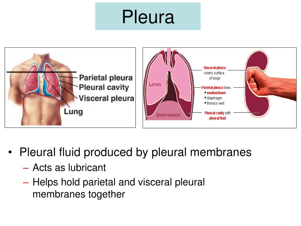
A camera can be inserted through a small incision in your chest during a procedure called a thoracoscopy. This can be done by inserting a small camera through your nose or mouth during a procedure called a bronchoscopy. Your doctor may want to get a closer look at the inside of your chest cavity. Thoracentesis can also be used to remove excess fluid from around the lungs.

If the fluid is infected, your doctor will want to address it quickly to prevent long-term damage. The fluid sample is analyzed for types of cells, chemical makeup, cultures and abnormal cells. This procedure, called thoracentesis, inserts a thin needle into the chest cavity, and removes a small amount of fluid. Your doctor may want to analyze your pleural fluid or tissue by taking a fluid sample or biopsy. They also assist in diagnosing underlying conditions, such as pulmonary embolism or lupus. This is another method that allows your doctor to examine the structures in and around the lungs.īlood tests help determine if you have an infection. Ultrasound uses sound waves to create images of your lungs.

Like a chest X-ray, a CT scan allows your doctor to look at images of your lungs, but in greater detail, using computer-generated images. The frequency of a positive TB-PCR result from the pleural fluid was significantly higher in the neutrophilic group than in. You may also need to undergo tests or procedures, such as the following: Chest X-rayĬhest X-rays provide an image of the inside of your lungs and allow your doctor to see whether fluid is present and how the lungs are functioning.Ĭomputerized Tomography (CT) Scan of the Chest Pleural fluid pH was significantly lower in the neutrophilic group than in the lymphocytic group (P0.019), while other biochemical markers, including ADA levels, were not significantly different between the two groups. Your doctor will perform a physical exam, listen to your chest and discuss your symptoms.


 0 kommentar(er)
